
A reference guide for storing laboratory solutions
Storing reconstituted peptide solutions: aliquots, freezing, containers and filters.
A peptide stock solution prepared by reconstitution for an assay or an analytical standard starts to change as soon as it is made. This guide covers how a laboratory stores that solution so its concentration stays measurable: aliquots, freezing and thawing, containers, filters and records. Every storage time quoted here is attributed to its published source.
For laboratory research use only. Not for human or veterinary use.
In this guide
Why does a peptide solution change faster than the powder?
A peptide stock solution is far less stable than the lyophilized powder it came from.
Bachem advises against storing peptides in solution, even sterile and oxygen-free solution, because they may slowly degrade, and GenScript calls their life in solution very limited.
A stored solution can change in three ways:
- Its chemistry changes. NIBSC, Bachem and GenScript each name sequences with cysteine, methionine, tryptophan, asparagine or glutamine among those with shorter lives in solution; their lists differ slightly.
- Its concentration changes without any chemistry. In recommendations on peptide standards for mass spectrometry, Hoofnagle and colleagues (2016) list evaporation and adsorption to surfaces among the main problems of extended storage, alongside microbial growth, which buffered solutions can support.
- Dilution makes both worse. In Thermo Fisher’s words, the lower the concentration, the more unstable the solution.
Where the sequence allows it, NIBSC and GenScript advise storing peptides in solution in a buffer at about pH 5 to 6, and NIBSC specifies a sterile one. A peptide whose isoelectric point lies near that range may be least soluble there, so solubility comes first; the charge and pH guide explains how to check.
Many peptides are also light-sensitive: Thermo Fisher advises keeping solutions out of direct light, and Hoofnagle and colleagues recommend amber or dark tubes for photosensitive sequences.
Surplus stock solution can be freeze-dried again, as GenScript and Thermo Fisher recommend for longer storage; the lyophilization guide covers what changes.
How long is a peptide stock solution stable?
The literature gives no single figure. Published guidance runs from a few weeks to about a year, depending on the source, the concentration and the temperature.
Hoofnagle’s figures describe mass spectrometry calibrators, typically in 5 to 30% acetonitrile with 0.1 to 1% formic acid, and their table places a frozen solution at high concentration in medium-term storage of three months to a year, depending on the peptide. Bachem’s “few weeks” is a general handling figure. Neither describes your solution.
The reliable answer is therefore a measured one: store the solution the way you intend to, and compare it with a fresh preparation at time points fixed in advance. The stability guide shows how to design that study.
- NIBSC
- Store aliquots at −20 °C or colder wherever possible, in a sterile buffer at about pH 5 to 6.
- Your laboratory
- The one figure that applies to your sequence, solvent, container and method.
These figures are attributed to their published sources. None of them is a stability claim for any GPC product or for a solution prepared from one.
How should a stock solution be divided into aliquots?
Aliquots let each analysis thaw only what it needs, so the rest of the stock is never frozen a second time. Bachem, NIBSC and GenScript all advise it, with frozen storage at −15 °C to −20 °C or colder depending on the source.
- 01
Keep the stock concentrated
Store the concentrated stock and make working dilutions from it as close to the time of analysis as possible, as Hoofnagle and colleagues recommend for calibrators. A dilute solution changes faster and loses a larger share of its peptide to the tube wall.
- 02
Size each aliquot to one run
No published source sets a minimum aliquot volume. Size each aliquot in µL to one method run and its replicate, so that no tube is thawed twice. Pipetting sets the practical floor: the concentration calculator asks for a precision check on transfers below 10 µL and calls for an intermediate dilution below 2 µL.
- 03
Match the tube to the volume
A small volume in a large tube has more wall per microlitre, and more of its peptide ends up on that wall. Kristensen and colleagues (2015) measured this at 2 µM in borosilicate glass: recovery of one peptide was 39% from 220 µL and 87% from 2 mL. Choose a tube close to the aliquot’s volume.
- 04
Choose the material with care
NIBSC calls glass and plastic vials generally satisfactory: polypropylene is strong and chemically inert, glass is the better choice where visibility matters, and plastics need care with organic solvents.
What do freezing, the freezer and air do to a stored solution?
Freezing slows chemistry, but it brings problems of its own. Four are worth planning for.
Thaw each aliquot once
Every source cited here advises against repeated freeze–thaw cycles, but the measured picture is more nuanced. In a controlled LC-MS study of 1 µM peptide aliquots stored at −80 °C, ten freeze–thaw cycles did not significantly change the mean peak area, but they left the peak areas more variable than any other condition did. The authors concluded that freeze–thaw cycles should be kept to a minimum.
Freezing concentrates what is dissolved
As ice forms, the peptide, salts and buffer are concentrated in the liquid that remains, and the pH can move with them. Gómez and colleagues found that sodium phosphate buffer at pH 7.4 reached pH 4.2 at −10 °C at 50 and 100 mM, and pH 5.2 at 8 mM. A thawed tube may not be uniform, so mix it gently and spin it briefly before pipetting.
Avoid automatic defrosting
A frost-free freezer warms periodically to melt its ice. NIBSC advises against frost-free freezers for peptides, because they “vary enormously in temperature during the frequent automatic defrosting”. Store frozen aliquots in a manual-defrost freezer at −20 °C or colder, or at −70 °C or colder for concentrated stocks held longer.
Limit air for oxidation-prone sequences
Cysteine, methionine and tryptophan oxidize readily, and Hoofnagle and colleagues report that the reaction speeds up during freeze–thaw cycles and at high pH. GenScript limits air by purging buffers with argon or nitrogen and keeping vials tightly capped, and Hoofnagle and colleagues add an inert blanket gas for particularly sensitive sequences. Bachem dissolves a peptide with a free cysteine in degassed acidic buffer, because thiols oxidize rapidly above pH 7. A reducing agent such as DTT is an option only if the method tolerates it.
Why does a dilute peptide solution lose peptide to its container?
Peptides in solution adsorb to tube walls, vials, pipette tips and cuvettes, and at high dilution the loss can be large.
A surface can hold only so much peptide. A container wall behaves a little like blotting paper under a drop of ink: it holds a roughly fixed amount. From a concentrated solution that amount is a small share; from a dilute one it can be most of what was there.
Hoofnagle and colleagues put the larger relative loss at low concentration down to the limited binding capacity of the wetted surface, and NIBSC warns that at high dilution, adsorption can grossly distort the measurements that follow.
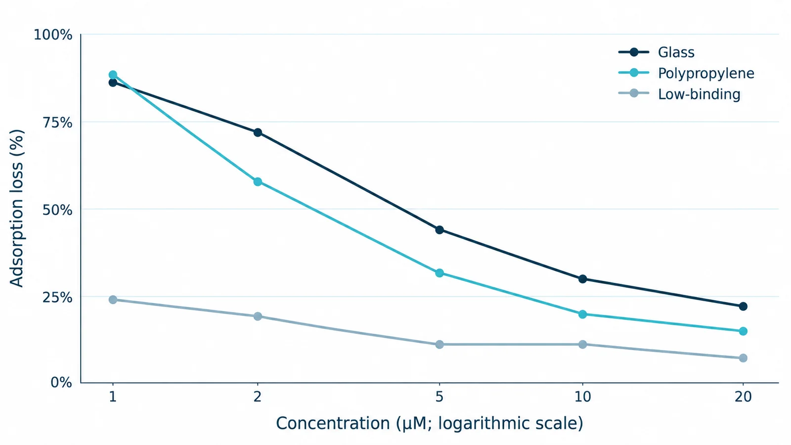
- GlassLoss: 86% at 1 µM; 22% at 20 µM.
- PolypropyleneLoss: 88% at 1 µM; 15% at 20 µM.
- Low-binding polypropyleneLoss: 24% at 1 µM; 7% at 20 µM.
Adsorption loss data table
| Container | 1 µM | 2 µM | 5 µM | 10 µM | 20 µM |
|---|---|---|---|---|---|
| Glass | 86% | 72% | 44% | 30% | 22% |
| Polypropylene | 88% | 58% | 32% | 20% | 15% |
| Low-binding polypropylene | 24% | 19% | 11% | 11% | 7% |
- 1 µM in glass or polypropylene
- Recovery of only 10 to 20% for three cationic peptides after 1 h in 220 µL at pH 7.4 (Kristensen and colleagues)
- 1 µM in low-binding tubes
- Recovery of 62 to 90% under the same conditions
- 20 µM in glass or polypropylene
- Mean recovery of about 78 to 85%
- Four transfers at 5 µM
- Recovery close to 0% after four successive glass or polypropylene containers, against 64 to 74% in low-binding tubes
- 0.2 µM against 1 µM
- In untreated glass vials over 15 h of hourly LC-MS runs, 9 of 50 peptides showed signal decay at 0.2 µM and none at 1 µM (Hoofnagle and colleagues)
Kristensen’s three peptides are strongly cationic and were measured at pH 7.4; the figures for low-binding tubes and at 20 µM are recalculated from the paper’s published dataset. Other sequences may lose more or less, so measure your own.
How do laboratories reduce loss to glass and plastic?
No container is best for every peptide, so the aim is to limit the loss and then measure what remains.
Glass or polypropylene: test, do not assume
Hoofnagle and colleagues describe basic residues binding the silanol groups of glass and non-polar residues binding polypropylene, and in their LC-MS comparison at 50 nM, polypropylene vials showed 1 unstable peptide against 13 and 14 in untreated and deactivated glass. Yet Goebel-Stengel and colleagues (2011), testing eight radiolabelled peptides of different charge and size, found no single best surface and no pattern by charge: recovery ranged from about 9% to 86%.
They help, but do not remove loss
At 1 µM, recovery of the three cationic peptides was 62 to 90% from low-binding polypropylene tubes, against 10 to 20% from glass and polypropylene, and an independent LC-MS study found more consistent replicate measurements with low-retention tubes than with standard microcentrifuge tubes. They are not a guarantee: at 2 µM in 2 mL, one peptide did no better in low-binding tubes than in glass or polypropylene.
BSA helps where the method allows it
In the Goebel-Stengel study, 1% BSA improved recovery for most peptides and never increased the loss, but BSA cannot be used in some mass spectrometry work, and Hoofnagle and colleagues find no consensus on which carrier to use or how much. A carrier also adds its own UV absorbance and peaks, so measure the stock first and add the carrier only to working dilutions whose method tolerates it.
Tips, transfers and pre-rinsing
Every tip, tube and transfer adds surface. Hoofnagle and colleagues advise a new tip for each dilution, pre-rinsed several times with the solution, and adding the peptide directly into the diluent rather than onto the tube wall. They also suggest washing tubes with the same solution first, while Kristensen and colleagues caution that a pre-saturated wall can later release peptide back into solution.
Measure the loss in your own containers
Hoofnagle and colleagues describe a direct check: transfer a solution from one vial to the next and analyze a small aliquot after each transfer. UV absorbance across a dilution series, or repeated LC-MS runs, shows loss to tubes, tips and vials in the same way. GPC’s Research Use Policy lists container and surface studies among its examples of permitted laboratory work.
Should a peptide stock be filtered, and through which membrane?
Filter only when the method calls for it, and then filter the concentrated stock once rather than each dilute working solution.
A membrane is one more surface, and a solution passing through it meets the membrane’s whole internal structure. Like a container wall, it holds only so much peptide, so the same loss is a smaller share of a concentrated solution. That is reasoning from the adsorption data above, not a measured peptide filter study.
Corning’s filtration guide sorts membrane pore sizes by purpose:
- 0.2 or 0.22 µm: routine laboratory sterilization of most media, buffers, biological fluids and gases.
- 0.45 µm or larger: clarification and prefiltration of solutions and solvents.
- 0.1 µm: mycoplasma removal.
GenScript presents 0.2 µm filtration as an option for removing possible bacterial contamination from a peptide solution. It does not establish sterility beyond the needs of a method, as the stock-solution guide explains.
A hazy stock is not a candidate for filtration. NIBSC notes that a solution that has gelled, stays hazy or carries a surface scum has probably not dissolved but is finely suspended, and filtering a suspension removes peptide instead of dissolving it. Diagnose it first with the troubleshooting guide.
The membrane rankings below come from protein data. For a peptide they are a starting point, and the recovery through your own membrane is the figure that counts: if the concentration matters to the method, measure it after filtration.
- Cellulose acetate and PES
- Very low protein binding (Corning). Surfactant-free cellulose acetate is the lowest; standard cellulose acetate carries under 1% wetting agent, which a little warm purified water rinses out.
- Regenerated cellulose
- Low protein binding, and Corning’s best choice for DMSO.
- Hydrophilic PVDF
- Grouped by KNAUER with cellulose acetate and PES for low protein binding. PVDF made for blotting binds protein strongly, so specify hydrophilic.
- Nylon
- Low to moderate binding (Corning), and KNAUER advises against it for protein-rich samples. It tolerates alcohols and DMSO better.
- Cellulose nitrate
- Very high binding (Corning), for solutions where protein binding is not a concern.
For a stock in DMSO, solvent compatibility decides first: regenerated cellulose tolerates DMSO, while PES has low chemical resistance. PTFE must be prewetted with a solvent such as ethanol before an aqueous solution will pass through it.
What should the label and the record say?
A stored aliquot is only as good as its label. Anyone who opens the freezer should be able to tell what is in the tube, how it was made and what has happened to it since.
Write these on the tube or its cryogenic label, and in the notebook:
- Identity: the compound and its lot number.
- Concentration: the value and its basis, weighed powder or corrected for net peptide content.
- Solvent: the solvent or buffer, and its pH.
- Preparation: the date and the preparer’s initials.
- Aliquot: its number and its volume in µL.
- Storage: the location and the temperature.
- History: a running count of freeze–thaw cycles, for any tube that goes back into the freezer.
Write with a solvent-resistant marker or on a printed cryogenic label, and keep the freezer’s temperature log with the notebook, so that any excursion is on record beside the aliquots it touched.
The stock-solution guide sets out the record made at preparation; these are the fields that storage adds.
When does a stored solution need measuring again?
Measure a stored solution again before it serves as a standard or calibrator whenever one of these applies.
- It looks different: haze, particles, a gel or a change of colour.
- The freezer had a temperature excursion.
- It has been stored longer than your own data cover.
- The method’s standards have drifted unexpectedly.
- The tube was frozen again after a thaw.
Measure the concentration, by UV absorbance or HPLC peak area against a freshly prepared reference, and the impurity profile: new or shifted peaks, identified by LC-MS with the help of the stability guide’s mass-shift table.
As a model, Hoofnagle and colleagues propose a minimum set of stability checks for mass spectrometry calibrators: 6 h and 24 h at 4 °C, 4 weeks at −70 °C, and one and two freeze–thaw cycles, each compared with time zero.
Whatever you find applies to that solution, stored that way, and sets no expiry date.
To have the material itself analyzed, a prepared solution is the wrong sample: the testing guide explains why laboratories need the original sealed vial.
Common questions about storing peptide solutions.
How long is a peptide stock solution stable at 4 °C, −20 °C or −80 °C?
The literature gives no single figure. Bachem says frozen solutions “may be kept for a few weeks” below −15 °C, while Hoofnagle and colleagues, writing on mass spectrometry standards, treat up to three months, liquid at 4 °C or frozen at −20 to −80 °C, as short-term storage for solutions of 1 to 100 µM, depending on the peptide. The only reliable figure is one your own method measures, and GPC makes no stability claim for any solution prepared from its products.
Does repeated freezing and thawing change a peptide solution?
Supplier guidance says to avoid it. In a controlled LC-MS study, ten freeze–thaw cycles of 1 µM peptide aliquots did not significantly change the mean peak area but made the peak areas more variable, and the authors concluded that freeze–thaw cycles should be kept to a minimum. Single-use aliquots, each thawed once, avoid the question.
Should a peptide stock be filtered at 0.22 µm or 0.45 µm?
Only when the method calls for it. A 0.2 or 0.22 µm membrane is the routine laboratory grade for removing bacteria, and 0.45 µm is for clarification and prefiltration. Filter the concentrated stock once, because a membrane holds only so much peptide, and never a hazy stock, which is a suspension that filtering strips of peptide.
How can you tell whether peptide was lost on a filter or changed?
Run the filtrate and an unfiltered portion of the same stock by HPLC, side by side. A smaller main peak with no new peaks points to loss on the membrane; new or shifted peaks point to chemical change, which the stability guide’s mass-shift table helps identify. If recovery is higher in later portions of filtrate than in the first, that pattern fits adsorption, because the membrane’s binding capacity fills up.
Why is a measured concentration lower than calculated in a dilute solution?
A surface holds a roughly fixed amount of peptide, so the share lost grows as the concentration falls, and every container, tip and transfer adds surface. Check the amount basis and the pipetting too, with the net-content guide and the concentration calculator, then keep the stock concentrated and make the final dilution in the analysis container just before the run.